What is Lamprobe?

As the greater mass of the American population ages, many minor skin abnormalities appear in the aging skin that may affect the aging baby boomers both physically and psychologically. The declining ozone layer has led to a dramatic increase in various skin disorders including sun damaged skin that may act as precursors to skin cancer. Treatment for these skin conditions is being sought after in today’s society because this population, besides wanting to look younger and more attractive as long as possible, are also very interested in preventative health care.
The skin changes that occur with aging, various skin abnormalities or lesions proliferate in certain ethnic populations. Minor skin conditions such as age spots, telangiestasia or skin tags are more commonly found in certain ethnic groups. For example, fibromas appear more frequently among the black population and telangiestasia (a.k.a. broken capillaries) are more visible on the cheek and nose of Caucasian skin. With age, multiple skin tags proliferate mainly on the neck, breasts and axillae, and in the skin folds in the obese. Oily skins suffer from asphyxiated pores with sebaceous secretions sometimes hardening on the skin’s surface, and no amount of manual extraction can release the suppressed sebum. Milias (a.k.a. whiteheads) are found on any skin type but occur more frequently in oily skins or very dry skins due to poor exfoliation. Among dark-skinned individuals (Fitzpatrick Skin Types III-VI) hyperpigmentation is a major source of concerns especially in aging.
Lamprobe helps remove these minor skin abnormalities.
Keratoses
Keratoses refer to the excess production of keratinized cells or hyperplasia. Keratoses are soft or hard keratinized cells and appear in different forms:
a) Seborrheic Keratosis: These are benign skin lesions that are small to large in size and are quite common during middle age or late years. They are not derived from sebum and actually originate in the epidermis. They appear warty, pigmented ( dirty yellow to black), scaly and somewhat flat in shape, sometimes with a cone-like projection and appear stuck on the skin. They are most common on the back, face, scalp and chest and differ from acitinic keratosis which are more erythematous (reddish) and scaly.
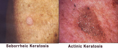
b) Acitinic Keratosis (“actinic” refers to sun) These are flat hyperpigmented lesions (a.k.a. liver spots) that appear dry, hard and rough in texture. They are commonly found in sun-exposed areas (face, arms, legs and back of hands) among middle-aged individuals, especially among those who tend to freckle easily e.g. blondes and red heads. Some eventually transform into squamous cell carcinoma. They differ from solar lentigo by appearing to be somewhat erythematous(reddish). These lesions are also commonly found among those individuals who are always in the sun and work in outdoor occupations such as gardeners. Repeated episodes of scaling occurs after sun exposure. They are different from seborrheic keratosis which are greasy, scaly and pigmented.
c) Fibromas : These are benign tumors that are either flat, raised, small or large. They either sit on the skin’s surface or are found attached by a short thin neck or peduncle. Pedunculated fibromas vary in size from pea-sized to larger ones and are soft tumours of normal to darker color. Sometimes they fall off by themselves.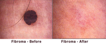
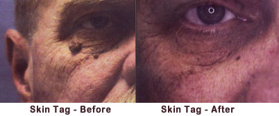
A skin tag is a small fibroma that appear either single or in multiple formation and sometimes referred to as an achrochordon. They are commonly found on the neck, breasts and axillae, and constant friction from necklaces or shirt collars contribute towards their formation in these areas. Sometimes they appear on the eyelids. They can become inflamed if they are repeatedly irritated when tissues are rubbed together in friction e.g. wearing chains on the neck or in the skin folds of the obese. After age 30, they proliferate and some individuals can have more than 200 or more on the breast or neck. This can pose psychological stress on some individuals who tend to wear scarves or high collars to hide them. Deratosis Papulosa Nigra are skin tags that are prevalent in Fitzpatrick Skin Types V to VI including African and Afro-Caribbean. These lesions appear in adolescence and increase with aging. They are darkly pigmented with no scales and believed to be genetically caused. They are commonly found on the face and neck, especially on the cheeks and orbital areas.
Sebaceous Lesions
These lesions are associated with sebum, the oily secretions of the sebaceous glands.
a) Milias (a.k.a. whitehead): These plugs of sebum area covered with layer/s of statum cornified cells and are commonly found on the facial area where there is poor skin exfoliation such as very oily or very dry skins. They can also occur during the healing of traumatic scars. Milias are commonly found on the forehead and cheeks, and when they are found under the eyes, they can be caused by eyeglasses that obstruct skin exfoliation. Our advice is to clean the eyeglasses regularly and use deep cleansing products and / or peeling masks to enhance skin exfoliation and thus prevent their formation.
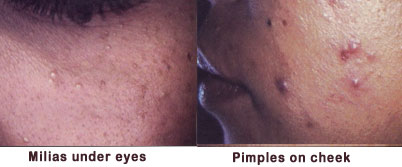
b) Pimples: These raised lesions are filled with pus and are common in acneic skins or skins with breakouts. Dirt, makeup and keratinized cells in the hair follicle pug the sebaceous duct. This blockage eventually leads to the rupture of the follicle into the upper layers of the dermis resulting in tissue inflammation. Phagocytes (white blood cells) rush to the area to protect the body and the resulting pus consists of dead bacteria, keratinized cells, free fatty acids and cholesterol. When the lesion is red and inflamed, it is called a papule and should not be touched. Many individuals find it irresistible and tend to squeeze the pimple causing infection, eventually leading to post-inflammatory hyper-pigmentation or scarring.
c) Sebaceous Cyst: This is a lesion associated with acneic skin and occurs when the overburdened sebaceous duct breaks down and leaks out into the deeper dermis leading to phagocytic action. A hardened wall forms around the infection and this appears on the skin’s surface as a hard, raised and painful lesion with no surface opening. The cyst also contains keratinized cells and is usually non-inflammatory unless it ruptures. If left alone, sometimes it can heal on its own. It occurs commonly on the scalp, face, neck or trunk and should be referred to a dermatologist to have them excised. The client should be advised to avoid touching it, stay on a healthy diet and refrain from eating inflammatory foods.
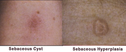
d) Sebaceous Hyperplasia: Chronic sun damage contributes towards these lesions resulting in enlarged sebaceous glands. They appear as a soft, yellowish papule with a cauliflower-like or doughnut-shaped appearance ranging in size from 2 to 3mm. They are usually solitary and appear on the forehead and cheeks, particularly on oily and asphyxiated skins. Manual extraction will not release any sebum and these lesions give a rough texture to the skin.
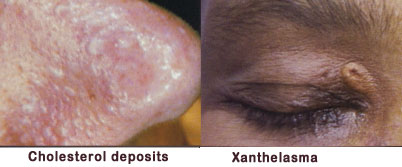
e) Xanthelasma (a.k.a. cholesterol deposits) These are soft yellowish plaques of lipids usually found in the periorbital (around eyes) area. They are very tiny to medium in size and some may be raised. Some individuals are predisposed towards them. They are believed to be related to abnormal lipid metabolism.
Vascular Lesions
They appear in different forms, an excessive amount giving the skin a reddish or “couperose” complexion. They may be hereditary or caused from any constant exposure to excessive blood stimulation such as alcohol, caffeine, hot drinks, physical exertion, excessive spicy foods, high blood pressure, drugs, sun exposure and high emotional individuals whose skins gets red or flushed easily. Vascular lesions are more noticeable in fair skinned individuals. (Fitzpatrick skin types I to II)
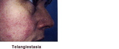
a) Telangiestasia ( a.k.a. broken capillary) These tiny, superficial dilated blood vessels appear as red wavy lines on the skin, particularly around the nose, cheeks and even the décolleté area. Constant blood stimulation causes the thin elastic walls of the capillaries to vasodilate eventually leading to breakage. Thin, blotchy skins as well as those who suffer from allergies are more likely to suffer from telangiestasia but they can also occur from skin trauma such as a fall. When they are small, bright red dots with radiating threads, they are called spider nevi. Compression of the central arteriole will blanch the lesion. Larger capillaries (a.k.a broken veins) on the legs are associated with the venous system and needs sclerotherapy, a medical procedure. They are commonly seen in women and may be associated with pregnancy, oral contraceptive use, and compression of tissues when women cross their legs regularly.
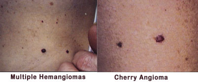
b) Hemangiomas ( a.k.a. angiomas, ruby points or blood spots) Hemangiomas are minor vascular abnormalities of the skin found on the face, neck chest or back. There are many types varying in depth (superficial to deep), clinical appearance and location. Cherry angiomas are bright red to purple dots of blood usually found on the upper trunk such as the neck area. In children, they are called strawberry hemangiomas and appear raised, red and soft with a strawberry-like lobule but usually disappear in adulthood.
 For treatment of